NMR Platform
NMR Platform
Nuclear Magnetic Resonance (NMR) technology at IPK
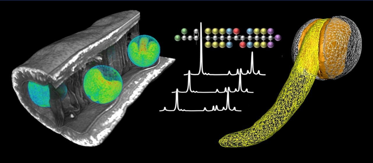
Nuclear Magnetic Resonance (NMR) technology at IPK is represented by a powerful technical platform made for high resolution functional imaging of plants. The goal and task of the NMR-platform at IPK is to enable non-invasive visualization and investigations of living plant and in particular seeds. Traits of interest can be analysed at many different levels – inner structure, biochemistry and dynamic - in the living specimens, and this makes the uniqueness of this technology.
The NMR-platform includes dedicated instrumentation, original methods and applications. The development of the NMR platform has been conducted by the Research Group Assimilate Allocation und NMR (AAN), headed by PD Dr. L. Borisjuk in the Department of Molecular Genetics (head Prof. T. Altmann); it was granted by the EFRE program (Europäischer Fonds für Regionale Entwicklung).
NMR operates in the radiofrequency region of electromagnetic spectra, and has been exploited to study various aspects of plant´s life (for review see Borisjuk et al., Plant Journal 2012). NMR instruments use magnetic fields, magnetic field gradients, and radio waves to characterise composition and/or generate images of the inner, hidden from human eyes structures and events. Few, if any, other analytical techniques addressing the physiology and development of living plants in their natural environment are as versatile as NMR (Ratcliffe, In Encyclopedia of Spectroscopy and Spectrometry, 2010).
In contrast to most of medical applications, which use standardised methods (pushbutton protocols), plant MRI is still challenged by relies by peculiarities of plant tissues. Thus, plant NMR-imaging still relies on original developments and technological adjustments (including hard and software).
The current NMR platform includes the following modules:
The vertical 9.4T superconducting magnet Bruker Avance III (Bruker BioSpin GmbH, Rheinstetten, Germany). This is a wide bore system (inner diameter 89 mm) operates at 400 MHz frequency and is capable of imaging. It is designed to generate three different magnetic fields: the first (B0) is established by a large static magnet, the second (Gx, Gy, Gz) by a gradient coil set which generates three switchable spatially varying orthogonal magnetic fields, and the third (B1) by a RF resonator which provides a temporal varying magnetic field orthogonal to B0 . The available set of RF coils for 1H NMR conventional imaging allows for analysis of specimens with resolution 30-60 mm. On the same scanner we also perform Magnetic Resonance Spectroscopic Imaging (MRSI), chemical exchange saturation transfer (CEST) MRI, and Chemical Shift Imaging (CSI) to generate spectroscopic information about the metabolic processes occurring in the plant specimen in addition to the image that is generated by 1H MRI.
Cryogenically-cooled double-resonant 1H-13C-probehead (Bruker BioSpin GmbH, Rheinstetten, Germany). This enables up to five-fold enhancement of the detection sensitivity in high-resolution NMR as compared to conventional probes. The sensitivity or signal-to-noise enhancement in the cryogenic probe is accomplished by lowering the temperature of the coil and the preamplifier. This does, however not affect the temperature of the specimen. Technology allows imaging with close to cellular resolution, and is used for advanced applications on seeds and other small plant organs/specimens. A prominent application example is the imaging and tracing of (13C-labeled) sucrose, representing the main phloem derived sugar feeding the seed.
High-end computer work station for three-dimensional (3D) modelling. This comes in combination with the software packages MATLAB and AMIRA, and is used for processing of 3D MRI data. Unlike a light microscopy image which is acquired in image space, the MRI image is acquired in Fourier space (also referred to as k-space), and the actual image then has to be reconstructed via a multidimensional inverse Fourier transformation. Unlike light microscopy images, NMR images are monochromatic, although colour coding can be added. Based on the 3D-datasets, derived from MRI or MRSI, we can generate 3D-models, allowing to visualize structures or calculate volumes of (sub-)organs and their dynamic changes during growth and development.
The Bruker Minispec MQ-60 TD NMR Analyzer (Bruker GmbH). This benchtop 1H-NMR instrument with 60 MHz operating frequency is equipped with a permanent 1.5 T strength magnetic field. Such types of instruments are widely used in medical research and quality control (food and pharmacy). We have achieved significant improvements in instrument performance by developing a novel, rapid, accurate procedure, designed to simultaneously quantify a number of basic seed traits without any seed destruction. Using this instrument, the procedure gives a high accuracy measurement of oil content, carbohydrates, water, and both fresh and dry weight of seeds. The non-invasive method requires a minimum of ~20mg biomass per sample, and thus enables to screen individual, intact seeds. (For more details see “A novel noninvasive procedure for high-throughput screening of major seed traits.” Rolletschek, H., Fuchs, J., Friedel, S., Börner, A., Todt, H., Jakob, P. M., L. Borisjuk, Plant Biotechnol. J. 13, 188–199, 2015). No imaging is possible using this instrument.
The automated sample delivery robot for MQ-60TD NMR, whichis designed for fully automated weighing of vials and vial transfer toward the mq60 NMR instrument. The robot was provided by LAIX Technologies (Langerwehe, Germany) and the software for robot operation was provided by Comicon GmbH (Hamburg, Germany). It consists of a xyz-picker arm, an electronic balance and two sample racks of 250 vials each. The vials have to be filled manually with appropriate amounts of seed, while all subsequent steps run fully automated. When combined with TD-NMR, a throughput of ~1,000 samples per day is achievable. We currently develop further procedures to widen the analytical spectrum and applicability of TD-NMR for seeds of various plant species.
Numerous new NMR imaging methods and measurement techniques have been developed by our research team, were published and implemented at IPK. For example, we apply MRI to obtain three-dimensional models of plant organs, internal structures and characterize metabolite distribution with high spatial resolution (Borisjuk et al., Plant Journal 2012; Munz et al., Biochimie 2016). Diligent adaption of NMR pulse sequences provides the means to visualise metabolic activities (Rolletschek et al., Plant Cell, 2011), to track the flow of sugars within a living seed (Melkus et al., Plant Biotech. J., 2011), to quantitatively map the distribution of storage lipids in seeds and fruits (Borisjuk et al., Progress in Lipid Research 2013; Sturtevant et al., Sci. Adv. 2020), to monitor the germination of seeds (Munz et al., New Phytol., 2017), to survey endosperm in mature seeds of distant crosses (Tikhenko et al., Comm. Biol., 2020), to comparatively analyse transgenic plants (Rolletschek et al., Plant Cell, 2020; Radchuk et al., 2021) and mutants (Meitzel et al., New Phytol., 2021). Various plant species and transgenic models can be safely investigated, e.g. oilseed rape, wheat, barley, soy, maize, pea, oat, rice, rye, tobacco, cactus, roses, sugar beet, Arabidopsis, Camelina, Jatropha.
The vertical 11.7T superconducting magnet AVANCE NEO (Bruker BioSpin GmbH, Rheinstetten, Germany). This is a super wide bore system operating at 500 MHz frequency and capable of imaging. A major advantage of the instrument to be acquired is that - due to the design of the instrument - not only seeds (or smaller objects of max. 2 cm size) but whole plants or complex plant organs (ears, pods, fruits, roots) can be investigated. This makes the measurements much more efficient and will significantly expand the application areas of NMR for plant research. Accompanied by appropriate method development, the IPK thus secures a decisive advantage in its core competence - research on crop plants and utilization of genetic diversity (biodiversity). The Super Wide Bore NMR device is currently the undisputed state of the art in the field of NMR-based imaging systems. The IPK has decided to expand the NMR technology platform at the IPK and to add another lighthouse project with international visibility to the existing infrastructure. Supported by the EFRE investment program, it is planned to establish this "super-wide-bore" NMR instrument at the IPK in 2022 in order to cover the future demands.
Contact:
Dr. Hardy Rolletschek
Phone: +49 39482 5-686
E-Mail: rolletschek[at]ipk-gatersleben.de

